Backgrounded membrane imaging (BMI)
Backgrounded membrane imaging (BMI) is an automated variation of the light microscopy compendial method, allowing the sizing, quantification and morphological characterization of micrometer-sized particles.
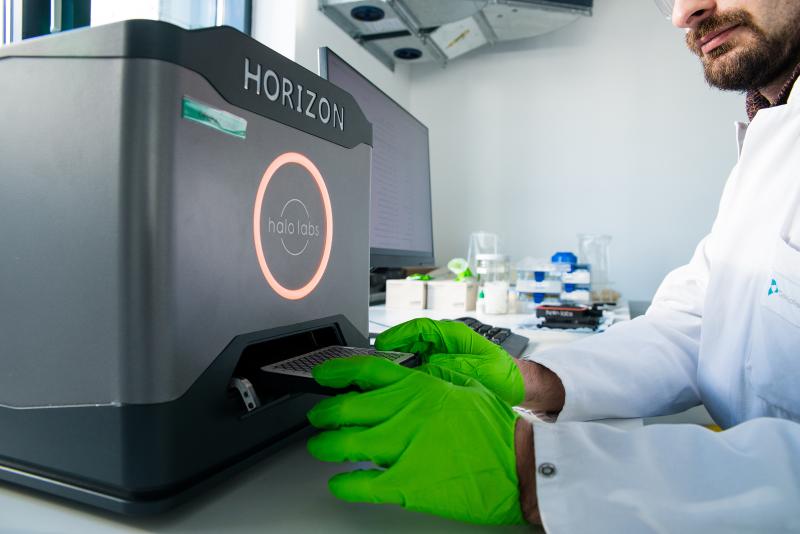
For BMI analysis, liquid samples are applied onto a 96-well filter plate and the suspended particles are filtered and dried on the filter membrane. Subsequently, a microscope camera captures bright-field images of the respective filter area. The digital images of the particles present on the filter are processed by background subtraction algorithms and image morphology analysis software that allows their quantification in size and count.
BMI is currently used as a research tool supporting more established particle characterization and quantification techniques, such as flow imaging microscopy [FIM] and light obscuration [LO]. The benefit of BMI is the low sample volume, the short measurement times owing to the well plate set-up and the supposedly higher image contrast compared to particles in suspension.
Need more information? Follow the links below and contact our experts with your questions today.
Contact us
Contact us
Need more information? We are happy to answer your questions!
Phone: +49 89 41 77 60 – 111
Mail: joerg.mueller@coriolis-pharma.com