Surface Plasmon Resonance (SPR)
Surface Plasmon Resonance (SPR) for Drug Product Characterization
In the field of pharmaceutical drug products, the activity of the drugs is usually based on a molecular interaction with specific receptors or target molecules in the human body. Surface Plasmon Resonance (SPR) allows reliable and detailed characterization of the drug product with respect to specificity and strength of the interaction to the target.
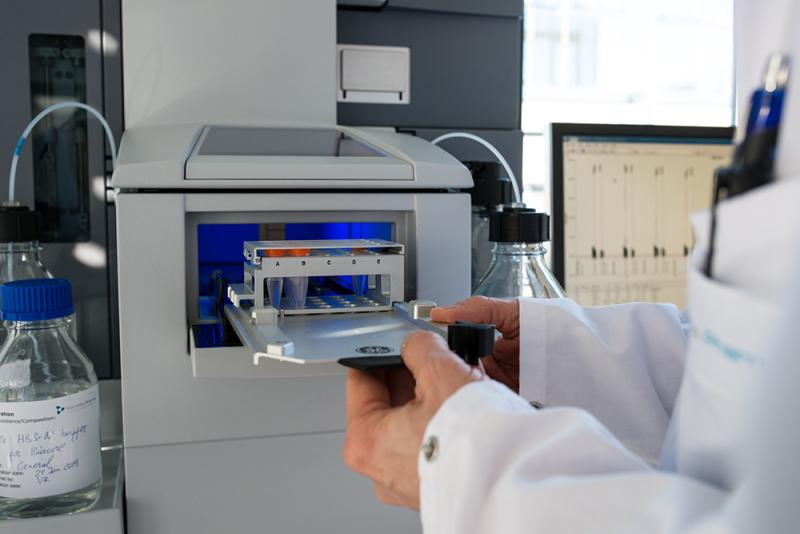
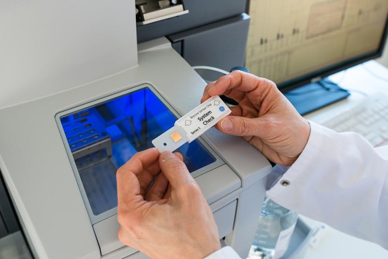
Biacore™ (Biacore™ T200 instrument) is a classic bioanalytical device based on the SPR principle to study interactions of any kind of molecule. To monitor the interaction between two molecules, one is attached to the sensor surface (dextran-coated surface of a sensor chip) and the other one is free in solution. The SPR optical detection system monitors the change in mass concentration in real time. These changes are recorded as sensorgrams. During sample injection, a positive response can be observed in the sensorgram, as the interacting partner in solution binds to the interaction partner attached to the sensor surface and therefore increases the mass concentration (association phase). The response decreases during dissociation.
Analytical Services with Biacore™:
- Concentration determination
- Absolute concentration (e.g. Protein A/G surface for IgG content)
- Active concentration (specific target surface) - Determination of the strength of an interaction pair; outcome are binding constants (kon = association rate constant, koff = dissociation rate constant; KD = Affinity)
- Receptor binding studies (e.g. Fcγ receptors)
- Feasibility studies for SPR-assay development
- Comparability and stability studies based on the potency assessment of samples.
Determination of Concentration
The direct binding assay is the most often used approach for concentration determination utilizing a calibration curve prepared by analyzing known concentrations of a reference sample under the same conditions. Drawback is that only the active portion of the sample is binding to the surface.
For the determination of the relative active concentration of e.g. IgG as drug product at first the absolute protein concentration is determined via a protein A/G surface. Protein A/G has a high affinity to the Fc part of IgG independent of the functionality of the Fab fragments. Stating the reference sample utilized for calibration curve to be 100% active the obtained active concentration of the sample can be set into relation to the absolute protein concentration. This is an important information for stability studies of IgGs as DP.
Fc-Receptor Binding Studies
Therapeutic antibodies and Fc-fusion proteins represent a significant segment of the biopharmaceutical industry and have resulted in substantial benefits to public health.
Antibodies bind to a specific target, but additionally to that they can regulate immune responses through interacting with Fc receptors (FcRs). The family of Fc receptors for IgG (FcγRs) is expressed on the surface of a variety of cells and an essential participant in many immune system effector functions. The receptors differ in their affinity and specificity for immunoglobulin subclasses. For example, antibodies of the IgG1 isotype have the potential to interact with all human Fcγ receptors (Nimmerjahn and Ravetch, 2008).
There are three major classes of FcγRs depending on the affinity of the receptor to IgG.
- Fcγ receptor I (CD64) can be found on monocytes and neutrophils. The most distinctive property of FcγRI is its relatively high affinity to IgG.
- Fcγ receptor II (CD32) allows phagocytosis and endocytosis of immune complexes and B cell activation and is a low affinity receptor.
- Fcγ receptor III (CD16) mediates antibody-dependent cellular cytotoxicity and is an intermediate affinity receptor.
Standardized Analysis at Coriolis Pharma
Characterization of the binding of therapeutic antibodies to His-tagged captured:
- human CD64
- human CD32a
- human CD16a
- human FcRn
Contact us
Contact us
Any questions about this method?
Please contact Dr. Jörg Müller for further information!
Phone: +49 89 41 77 60 – 111
Mail: joerg.mueller@coriolis-pharma.com